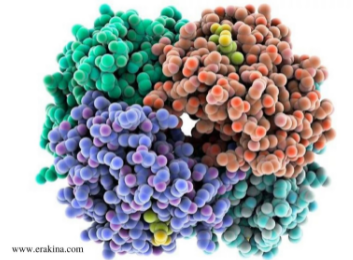
Complement proteins are proteins that assist the immune system in responding to the incoming pathogens by the process of opsonization.
Complement proteins are responsible for destroying the harmful pathogen by destroying their outer cell membrane or by allowing them to attract macrophages. These macrophages are the phagocytic cells, and this process of tagging the pathogens to get destroyed by phagocytes is called opsonization. Additionally, some complement proteins stimulate cells to release histamines, causing inflammation and directing the phagocytic cells to the infection site. The complement proteins are involved in a complement system that also fights infections and develops immune diseases. Three pathways activate the complement proteins; the antibody-dependent pathway (classical), the antibody-independent pathway (alternative), and the lectin pathway (Mannose binding-lectin – MBL). The complement proteins are indicated as C (complement) with a number (a component), such as C3. The improper regulation of the complement system can lead to potential tumors.
What are Complement Proteins?
Complement proteins are plasma proteins that are activated when the immune response is initiated upon the entrance of a pathogen into the body. The complement proteins tag the pathogenic cells by attracting the macrophages (type of white blood cells) onto these pathogens, called opsonization. This causes inflammation by releasing histamines in the surrounding area of pathogens present, attracting the macrophages (phagocytic cells) to the pathogens. The pathogen can either directly or indirectly (pathogen-bound antibody) is detected and tagged by the complement proteins for the phagocytic cells to destroy the pathogens.
The complement proteins that tag the pathogens are opsonins bound to the pathogenic cells, causing cell lysis, thus inducing phagocytosis. Phagocytosis is the process of releasing phagocytes when attached to a cell to phagocytose (ingesting or engulfing) the cell. The phagocytosis will kill the pathogenic cell, thereby eliminating the pathogens hence reducing the infection. Additionally, the complement system plays a role in clumping the pathogenic cells together, called agglutination. There are around 30 complement proteins found in the tissue fluids. These proteins are initially inactive, but in the presence of pathogens, the response from the immune system will activate these proteins. Understanding the complement system offers perception in tumor immunology for cancer diagnosis and treatment by regulating the complement system. An imbalance in the complement system leads to various autoimmune disorders and tumors.
Three Pathways of Complement System
The complement system is activated through three different pathways; the antibody-dependent pathway (classical), the antibody-independent pathway (alternative), and the lectin pathway (Mannose binding-lectin – MBL). A few examples of complement protein components are C2 through C9. C3 is one of the complement proteins that indicate the immune system’s response from various parts in the presence of harmful pathogens. The three pathways involved in forming C3 are later converted into C3a and C3b. This C3 convertase leads to the formation of MAC(Membrance attack complex), followed by cell lysis.
Classical Pathway
When a pathogen enters the body, the pathogenic cell contains antigens. In the presence of a pathogen, immune complexes such as IgG or IgM are produced, which bind to these pathogens or other foreign antigens in the classical pathway. The classical pathway is activated when the C1 protein binds to the surface of the pathogenic cell. A Y-shaped antibody contains two antigen-binding sites and one stalk Fc region. The first complement protein component to respond is C1, composed of C1q, C1r, and C1s (two proteases). Once the immune system detects the presence of foreign antigens, antibodies are released that bind to these antigens; thus occupying the two antigen-binding sites and forming an antibody-antigen complex. The C1 protein binds to the Fc region of the antibodies. Then C1 proteases cleave C4 protein into C4a and C4b, of which C4b binds to the cell surface of the pathogen. The cleavage of C2 follows this step into C2a and C2b, in which C2a binds to the C4b. This C4bC2a complex is the convertase of C3. This C3 is cleaved by C3 convertase to form C5 convertase completing the classical pathway.
Alternative Pathway
The first response of the immune system activates an alternative pathway. C3 protein is the most abundant in the complement system. This pathway commences by activating the C3 protein that requires two factors; B and D. At the beginning, spontaneous hydrolysis of C3 occurs that opens a portion in the pathogenic cell where factor B binds to C3(H2O)molecule. Factor D cleaves the factor B into a larger fragment (Bb – bound to the C3 molecule) and a smaller fragment (Ba). C3(H2O)Bb complex cleaves C3 into multiple C3 convertases. These C3 convertases (C3b) bind to the pathogenic cell surface.
Lectin Pathway
The lectin pathway is similar to the classical pathways where the lectins bind to specific carbohydrates on the pathogenic cell surface. This pathway is activated when the binding of MBL to mannose residues present on the pathogenic cell surface occurs. The serine proteases associated with MBL, MASP-1, and MASP-2 are activated to initiate cleavage of C4 and C2 proteins to produce C3 convertase, hence similar to the classical pathway. MASP-2 cleaves C4 into C4b, which binds to the cell surface, and C4a, released. MASP-2 also cleaves C2 into C2a and C2b. C2a binds to C4b to form the C4bC2a complex, a C3 convertase.


